
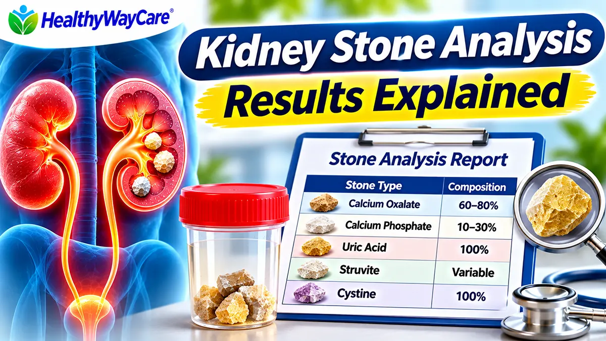
Kidney stone analysis results identify the minerals in a stone you passed or had removed. A laboratory may report calcium oxalate, calcium phosphate, uric acid, struvite, cystine, or a mixture. Knowing the composition helps your clinician decide which follow-up tests and prevention steps make sense. A result is not, by itself, a diagnosis of why the stone formed or a prediction that another stone is inevitable.
Quick answer: Calcium-containing stones are most common, but different types have different causes and prevention strategies. If your report lists percentages, they describe the composition of the submitted specimen—not your chance of another stone. Review the result with a urologist, especially if you have recurrent stones, infection, kidney disease, or a family history of stones.
Medical review note: This educational guide summarizes major urology guidance and is not individualized medical advice. Laboratory terminology and clinical interpretation can vary.
Table of Contents
Kidney Stone Analysis Results: Quick Reference Chart
| Report finding | What it generally indicates | What to discuss with your clinician |
|---|---|---|
| Calcium oxalate | A common calcium-based stone; may be monohydrate or dihydrate | Urine volume, sodium, calcium, oxalate and citrate |
| Calcium phosphate | A calcium stone often associated with higher urine pH in some patients | Urine pH, calcium, possible underlying conditions |
| Uric acid | A stone associated particularly with persistently acidic urine | Urine pH, metabolic health, diet and alkalinization |
| Struvite | An infection-associated stone containing magnesium ammonium phosphate | Urine culture, infection management, complete stone clearance |
| Cystine | An uncommon stone associated with inherited cystinuria | Specialist follow-up, urine cystine, fluid and pH goals |
| Mixed composition | More than one mineral identified in the submitted sample | Which components dominate and whether more testing is needed |
This chart is a starting point, not a treatment algorithm. For example, a calcium phosphate result does not prove renal tubular acidosis, and a uric acid result does not mean a person must have high blood uric acid. The clinical picture matters.
What Is a Kidney Stone Analysis Test?
A kidney stone analysis is a laboratory examination of a physical stone or stone fragment. It differs from a urinalysis, which examines urine, and from imaging, which helps locate a stone. A clinician may request analysis after a stone passes spontaneously or after a procedure such as ureteroscopy or percutaneous stone removal. The laboratory identifies crystalline components and may report one or more minerals.
The European Association of Urology (EAU) recommends analysis of stones in first-time stone formers using a validated technique. Repeat analysis can be valuable if a stone recurs despite preventive treatment, if another stone forms soon after complete clearance, or if recurrence occurs after a long stone-free interval. Stone composition can change over time, so an old report may not describe every future stone.
In the United States, your urologist may order stone analysis through a hospital or reference laboratory. Reports vary: some give the predominant mineral, others list several percentages, and some identify a specific crystal form such as calcium oxalate monohydrate. Your provider should interpret those findings together with your symptoms, imaging, blood work and urine chemistry.
How Do Laboratories Identify Kidney Stone Composition?
Two well-established methods are infrared spectroscopy and X-ray diffraction. These techniques identify mineral structure rather than simply measuring chemicals dissolved from a specimen. EAU guidance favors validated mineral-identification methods and considers older wet-chemical approaches less reliable for modern stone analysis.
The report may mention calcium oxalate monohydrate (whewellite), calcium oxalate dihydrate (weddellite), hydroxyapatite, brushite or magnesium ammonium phosphate (struvite). These names sound technical, but they provide more detail about the crystal composition. They do not necessarily tell you when the stone formed, how long it was present, or whether a current obstruction has resolved.
How to Collect a Passed Kidney Stone for Testing
- Ask your healthcare team for a urine strainer or a clean collection method if they want you to capture a stone.
- Strain urine as directed while waiting for passage; tiny fragments can be easy to miss.
- If a stone is recovered, follow the laboratory’s specific packaging and drying instructions. Do not add household chemicals or preservatives unless instructed.
- Label the specimen as requested and bring it to the clinic or laboratory with the order.
- Tell your clinician if you did not capture the stone. Imaging or other testing may still be needed to confirm passage.
Do not delay urgent evaluation in an attempt to collect a specimen. Fever or chills with suspected urinary obstruction, inability to urinate, uncontrolled pain or persistent vomiting can require emergency assessment. A stone analysis cannot diagnose an acute infection or determine whether a kidney is currently blocked.
How to Read Kidney Stone Analysis Results: A Sample Report
Consider this illustrative example, not a real patient report: “Calcium oxalate monohydrate 80%; calcium phosphate (apatite) 20%.” This means the submitted material was identified as predominantly calcium oxalate, with a smaller calcium phosphate component. It does not mean that 80% of the kidney is affected or that you have an 80% recurrence risk.
Some labs report a single component at 100%, while others provide multiple components. A specimen containing fragments from more than one stone can complicate interpretation. If a report says “unidentified material,” “insufficient specimen,” or “not a calculus,” contact the ordering clinician about whether further analysis is possible. The appearance or color of a stone alone is not a reliable substitute for laboratory identification.
| Example wording | Plain-English meaning | What it does NOT prove |
|---|---|---|
| 100% calcium oxalate monohydrate | The submitted stone material was identified as this crystal type | That your blood calcium is high |
| 70% calcium oxalate, 30% calcium phosphate | The sample contains two identified mineral components | A 70% chance of future stones |
| Predominantly uric acid | Uric acid is the major identified component | That serum uric acid must be elevated |
| Struvite detected | An infection-related mineral is present | That an active infection is definitely present today |
| Cystine identified | A finding strongly suggestive of cystinuria | That family members automatically have the disorder |
Calcium Oxalate Stone Results Explained
Calcium oxalate is the most common mineral found in kidney stones. Reports may distinguish monohydrate and dihydrate forms. These distinctions can be relevant to stone properties and specialist decisions, but prevention depends on more than crystal type. A calcium oxalate stone can develop when urine becomes concentrated or when urinary calcium, oxalate, citrate and other factors favor crystallization.
One of the biggest misconceptions is that a calcium stone means you should stop eating calcium. For many calcium oxalate stone formers, maintaining an appropriate amount of dietary calcium with meals is helpful because calcium in the digestive tract binds some dietary oxalate. Unnecessarily restricting calcium may harm bone health and can increase oxalate absorption. Discuss your dietary needs with your clinician, particularly if you have kidney disease or take supplements.
Sodium intake also matters. High sodium intake can increase urinary calcium excretion in susceptible people. A 24-hour urine collection may reveal elevated urine calcium, high oxalate, low citrate or low urine volume, each of which suggests different prevention priorities. Blanket avoidance of every oxalate-containing food is usually less useful than individualized advice based on your risk profile.
Calcium Phosphate Stone Results Explained
Calcium phosphate stones include minerals such as hydroxyapatite and brushite. They are distinct from calcium oxalate stones even though both contain calcium. Higher urinary pH can favor some calcium phosphate crystallization, and certain patients may have an underlying condition such as distal renal tubular acidosis or hyperparathyroidism. The stone report alone cannot diagnose either condition.
Because urinary pH affects different stone types in different ways, self-prescribing alkalinizing supplements is not a safe universal strategy. Raising urine pH may help prevent uric acid stones but may increase calcium phosphate supersaturation in some circumstances. Your clinician may review urine pH trends, blood bicarbonate, calcium, parathyroid-related testing when indicated, and 24-hour urine findings before selecting treatment.
If your report mentions brushite, ask whether it changes your follow-up plan. Stone mineralogy can sometimes influence procedural planning and recurrence assessment, but a report must be considered alongside imaging and clinical history.
Uric Acid Stone Results Explained
Uric acid stones often form when urine remains too acidic. They can occur even when blood uric acid is not markedly elevated. Factors such as metabolic syndrome, type 2 diabetes, dietary patterns and urine volume can influence risk. Unlike many calcium stones, uric acid stones may be poorly visible on standard X-rays, although CT imaging can identify them.
Clinicians may recommend increasing urine volume and, when appropriate, medication to raise urine pH to a carefully selected target. Some uric acid stones can dissolve with supervised urine alkalinization, but not all stones are uric acid and a mixed stone may not dissolve completely. Do not assume a stone is uric acid based only on urine color, pain pattern or an internet photo.
Reducing excessive purine-rich animal foods may be part of a prevention plan, but medication choices and pH targets require individualized assessment. If you have gout, diabetes, chronic kidney disease or take medications that affect potassium, tell your clinician before starting any stone-prevention products.
Struvite Stone Results Explained
Struvite is magnesium ammonium phosphate and is classically associated with urinary infections caused by certain urease-producing bacteria. Infection-related stones can enlarge and sometimes form branching staghorn stones. A report showing struvite should prompt discussion of urine culture, imaging and whether any stone material remains.
Treatment usually focuses on managing the infection and achieving appropriate stone clearance, because retained fragments can contribute to ongoing problems. Antibiotics alone do not necessarily eliminate a stone. Do not treat a struvite finding with home remedies or assume that symptoms have resolved because the pain has temporarily improved.
A struvite component does not prove you currently have a urinary tract infection; the clinician may need a culture and examination. Conversely, fever or chills with a suspected obstructing stone require urgent medical evaluation regardless of what an earlier stone analysis showed.
Cystine Stone Results Explained
Cystine stones occur in people with cystinuria, an inherited condition that causes excessive cystine in urine. They are less common than calcium stones and often require specialized long-term prevention. A cystine result should lead to a discussion about confirming the diagnosis, assessing family history and arranging appropriate urology or nephrology follow-up.
Prevention may involve very high urine output goals, dietary sodium reduction, carefully monitored urine alkalinization and sometimes cystine-binding medications. These measures are not appropriate for everyone and should be guided by a specialist, particularly in children, pregnancy or people with impaired kidney function.
Because cystinuria is genetic, relatives may benefit from individualized counseling or testing recommendations. A family history alone is not enough to diagnose the disorder, and genetic or biochemical evaluation may be considered depending on circumstances.
Mixed Kidney Stone Composition: Why Two Minerals May Appear
A mixed report is common enough that it should not be treated as a laboratory error. A stone may contain a core of one mineral and an outer layer of another, or the submitted specimen may contain multiple mineral components. Different conditions can influence mineral deposition at different times. The laboratory may report components by proportion, sometimes rounded to whole percentages.
Your clinician may focus on the dominant component while also considering the clinical importance of a smaller component. For instance, a minor struvite component can raise questions about infection, and a calcium phosphate component may make urine pH especially relevant. The percentages alone do not establish which condition caused the stone.
If the composition changes from one episode to another, that is a reason to revisit prevention rather than assume the first plan still covers every risk. Repeat stone analysis is particularly useful when new stones form despite treatment.
Stone Type vs. Possible Risk Factors and Prevention
| Stone type | Factors commonly assessed | Prevention approach to discuss |
|---|---|---|
| Calcium oxalate | Low urine volume; urinary calcium, oxalate and citrate; sodium intake | Fluids, appropriate dietary calcium, sodium reduction and targeted therapy |
| Calcium phosphate | Urine pH, urinary calcium and relevant metabolic conditions | Individualized pH assessment and calcium-stone risk reduction |
| Uric acid | Low urine pH, metabolic factors, urine volume | Hydration and clinician-supervised urine alkalinization |
| Struvite | Infection, urine culture and residual stone fragments | Infection treatment and evaluation for stone clearance |
| Cystine | Cystinuria, cystine concentration, urine pH and volume | Specialist-directed fluids, pH management and sometimes medication |
What Tests May Be Needed After a Stone Analysis?
Stone analysis is one piece of a broader evaluation. A first-time stone former may need a medical and dietary history, urinalysis and blood tests. People with recurrent stones, high-risk features or a strong desire to prevent recurrence may benefit from more detailed metabolic testing. The 2026 American Urological Association (AUA) guideline describes 24-hour urine evaluation as a way to identify modifiable factors in appropriate patients.
A 24-hour urine panel may include total volume, pH, calcium, oxalate, citrate, uric acid, sodium, potassium, creatinine and additional measures. One or more collections may be requested depending on clinical circumstances and laboratory protocol. These values help explain why a stone may have formed; they do not replace the direct analysis of the stone itself.
For a step-by-step overview, see our separate HealthyWayCare guide to 24-hour urine testing once it is published. Your clinician may also use imaging to look for remaining stones or obstruction, and a urine culture if infection is suspected.
How to Prevent Another Kidney Stone After Getting Your Results
The strongest prevention plan starts with the actual stone type and your individual risk factors. Many stone formers benefit from sufficient fluid intake to maintain a generous urine volume, but the appropriate target varies. People with heart failure, advanced kidney disease or prescribed fluid restrictions should follow their treating clinician’s instructions instead of generic high-fluid advice.
For many adults without a fluid restriction, urology guidance commonly aims for roughly 2 to 2.5 liters of urine output per day. Achieving that may require drinking more than that amount, particularly during hot weather or physical activity. Your provider can help set a target based on 24-hour urine results. Water is usually a practical first choice; our guide to drinks for kidney stone prevention discusses beverage options and limitations.
Dietary changes are not identical for all stones. People with calcium stones should not automatically eliminate dairy. Sodium reduction can be helpful for calcium stone prevention, while purine moderation may be relevant for some uric acid stone formers. High-dose vitamin C or unreviewed supplements can create problems for some people. A registered dietitian familiar with kidney stones can help make a plan that is realistic and nutritionally complete.
Follow-up matters because risk factors can change. Depending on the initial results and the prevention plan, clinicians may repeat urine testing to see whether urine volume, citrate, calcium, oxalate or pH has improved. Imaging follow-up may be needed even if you have no pain, because some stones cause few symptoms.
When to Seek Urgent Medical Care
Stone composition is useful for long-term planning, but it does not make current symptoms safe to ignore. Seek urgent medical assessment for fever or shaking chills with suspected stone symptoms, inability to urinate, uncontrolled severe pain, persistent vomiting or signs of serious illness. An infected, obstructed urinary system can become a medical emergency. Sudden severe testicular pain also requires immediate emergency assessment for other possible causes.
If you are experiencing flank or groin pain and are unsure of the cause, see our kidney stone pain location chart for a general symptom overview. Pain location alone cannot confirm a stone, its composition, or whether it has passed.
Questions to Ask Your Urologist About the Report
- Which mineral makes up most of my stone, and are there clinically important secondary components?
- Does this result suggest I need blood tests, a urine culture, or a 24-hour urine collection?
- Could my medications, supplements, diet, medical history or family history contribute to this stone type?
- Do I still have stones or fragments on imaging, and how will passage or clearance be confirmed?
- What fluid, sodium, calcium and other dietary targets apply to me specifically?
- Should I repeat stone analysis if another stone occurs, and when should I return for follow-up?
Frequently Asked Questions About Kidney Stone Analysis Results
1. What does a kidney stone analysis report show?
It identifies minerals in the submitted stone or fragment. A report may list calcium oxalate, calcium phosphate, uric acid, struvite, cystine or a combination. The report does not directly measure your risk of future stones or confirm whether the urinary tract is currently blocked.
2. What does 80% calcium oxalate and 20% calcium phosphate mean?
It describes the mineral composition identified in the submitted sample. It does not mean an 80% chance of recurrence. Your urologist may consider both components when deciding whether to check urine calcium, oxalate, citrate and pH.
3. Is calcium oxalate the most common kidney stone type?
Yes. Calcium stones are the most common broad category, and calcium oxalate is more common than calcium phosphate. Finding calcium oxalate does not automatically mean you consume too much dietary calcium.
4. Can a kidney stone analysis tell whether I have an infection?
A struvite component can suggest an infection-related stone, but a stone analysis cannot establish whether you currently have an infection. Symptoms, urine testing and culture may be needed. Fever with suspected obstruction requires urgent evaluation.
5. Can kidney stone composition change over time?
Yes. Some people form stones with different compositions at different times. That is one reason repeat analysis may be appropriate after recurrence, particularly when stones form despite preventive treatment.
6. Is stone analysis the same as a 24-hour urine test?
No. Stone analysis examines the physical stone; a 24-hour urine test measures substances and characteristics of urine that influence stone formation. They provide complementary information for prevention planning.
7. Can I identify my kidney stone type by its color or shape?
No. Appearance alone is unreliable. A validated laboratory technique such as infrared spectroscopy or X-ray diffraction is a better way to identify stone minerals.
8. What should I do if my report says uric acid or cystine?
Discuss the result with your urologist. Uric acid stones often call for assessment of urine pH and possible supervised alkalinization; cystine stones warrant evaluation for cystinuria and specialist-directed prevention. Do not start medications based only on the report.
Key Takeaways
Kidney stone analysis results provide a mineral fingerprint of a submitted stone. Calcium oxalate and calcium phosphate stones differ from uric acid, infection-associated struvite and inherited cystine stones. Mixed results are possible, and composition percentages are not recurrence probabilities. The best next step is to combine the report with medical history, urine chemistry, blood tests and imaging as appropriate, then create a prevention plan tailored to the stone type.
Medical Sources and Further Reading
- NIDDK: Definition & Facts for Kidney Stones — descriptions of major stone types.
- European Association of Urology: Urolithiasis Guidelines (2026) — stone composition and validated analysis methods.
- EAU: Metabolic Evaluation and Recurrence Prevention — stone-specific evaluation and preventive measures.
- American Urological Association: Medical Management of Kidney Stones Guideline (2026), Part I — evaluation and 24-hour urine testing.
About the Author
Yamanandan Reddy is a health content writer and frontend developer who develops research-based educational content for HealthyWayCare. This article summarizes published medical resources and does not claim that the author is a physician or that the article has undergone clinician review.
Medical Disclaimer
HealthyWayCare provides educational information only. This article is not a substitute for medical diagnosis, individualized treatment or emergency care. Ask a licensed healthcare professional to interpret your own report. The content should undergo independent clinical review before publication.