
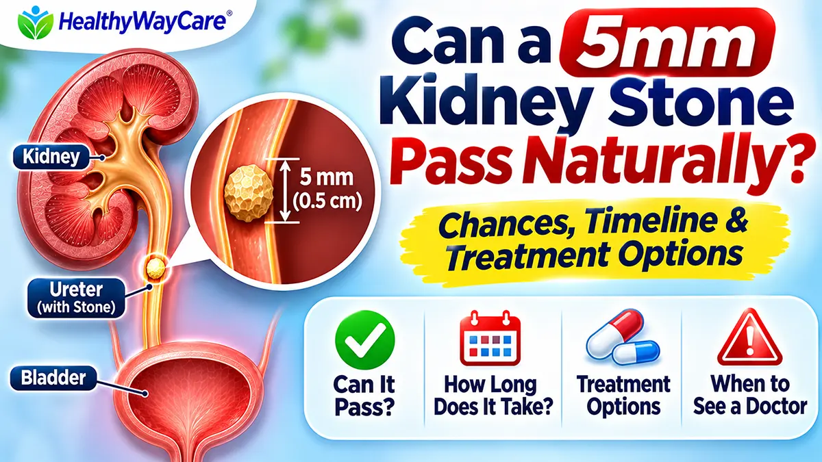
Can a 5mm kidney stone pass naturally? Yes. Many 5-millimeter stones in the ureter pass without surgery, but passage is not guaranteed. Where the stone sits, whether it blocks urine, your kidney function, symptoms, and signs of infection matter as much as its measured size. A 5 mm stone still inside the kidney is a different situation from a 5 mm stone moving through the ureter.
If a scan report says “5 mm calculus,” it is reasonable to ask two questions: Where exactly is it? and Is it causing obstruction? Those answers help determine whether observation is safe, how follow-up should work, and when a procedure might be preferable. This guide explains the evidence without promising a particular outcome or encouraging you to wait through dangerous symptoms.
Urgent warning: A suspected stone with fever or shaking chills, inability to pass urine, uncontrolled pain, repeated vomiting, or feeling seriously unwell needs urgent medical assessment. Infection behind a blocked kidney is an emergency, even when the stone is small.
Table of Contents
Quick Answer: What Are the Chances a 5mm Kidney Stone Will Pass?
The 2026 European Association of Urology (EAU) guideline summarizes studies in which approximately 62% of ureteral stones measuring 5 mm or larger passed spontaneously. This is a broad pooled category, not a 62% probability for an individual 5 mm stone. In the same evidence summary, passage varied by ureteral location: approximately 49–52% for upper, 58–70% for middle, and 68–83% for lower ureteral stones. These location estimates combine different stone sizes and patient groups. They should not be interpreted as exact personalized odds.
For someone with a confirmed 5 mm ureteral stone, stable kidney function, no infection, tolerable symptoms, and reliable follow-up, a clinician may recommend a monitored trial of passage. Someone with persistent obstruction, reduced kidney function, uncontrolled symptoms, or infection may need earlier intervention. The decision is medical, not simply a contest between “natural” and “surgical” treatment.
How Big Is a 5mm Kidney Stone?
Five millimeters is half a centimeter, roughly the width of a small pencil eraser. It sounds tiny, but the ureter is a narrow, muscular tube, and a stone can catch at naturally tighter points. The measurement on an imaging report is usually an estimate of a dimension of the stone; its shape and orientation also affect how it moves. A stone that looks round on one scan slice may be longer in another direction.
A 5 mm stone may cause little discomfort if it remains in the kidney without blocking drainage. The same stone can cause intense, wave-like pain after entering the ureter. Pain severity does not reliably reveal whether a stone is 3 mm, 5 mm, or 8 mm. A small stone can obstruct, while a larger nonobstructing kidney stone can sometimes be painless.
Why Stone Location Matters More Than People Expect
A stone begins in the kidney. If it enters the ureter, it must travel toward the bladder before leaving the body through the urethra. A stone high in the ureter has a longer path and may be less likely to pass spontaneously than a similar-sized stone near the bladder. The EAU guideline reports higher passage rates for distal than proximal ureteral stones across the studied groups.
5mm Kidney Stone Passage by Location
| Location on scan | What it means | Why it changes the plan |
|---|---|---|
| Inside kidney, not obstructing | Stone has not entered the ureter | May be monitored; ureteral passage percentages do not directly apply |
| Upper/proximal ureter | Near the kidney | Typically a longer route; passage less likely than for distal stones |
| Middle ureter | Partway toward the bladder | May pass, depending on obstruction and symptoms |
| Lower/distal ureter | Close to the bladder | Generally more favorable passage location |
| At ureterovesical junction (UVJ) | Where ureter enters bladder | May cause urgency; follow-up still needed to prove passage |
The table describes general patterns, not a diagnosis. A scan may also mention hydronephrosis, meaning swelling of the kidney from backed-up urine. Mild swelling can accompany a passing stone, but persistent or severe obstruction requires clinical attention. Ask your clinician to explain both the stone position and the degree of obstruction.
3mm vs 4mm vs 5mm vs 6mm Kidney Stones: What Changes?
| Approximate stone width | General passage pattern | Important caution |
|---|---|---|
| 3 mm | Often passes if in the ureter | Infection or obstruction can still make it urgent |
| 4 mm | Many pass without a procedure | Location and follow-up still matter |
| 5 mm | Passage is possible, but less predictable | A borderline size; avoid relying on a single percentage |
| 6 mm | Spontaneous passage becomes less likely | A urologist may discuss medicines or procedures |
| 8–10 mm | Often needs closer specialist consideration | Individual anatomy and location affect choices |
These are qualitative comparisons rather than fabricated stone-size-specific success rates. Research studies use different imaging measurements, follow-up periods, and definitions of successful passage. The EAU guideline distinguishes stones smaller than 5 mm from those 5 mm and larger, which makes a precisely measured 5 mm stone a threshold case rather than automatically a “small stone that will pass.”
How Long Does It Take for a 5mm Kidney Stone to Pass?
For a stone that does pass, the process may take days to several weeks. In studies summarized by the EAU, the average reported time to expulsion was around 17 days, with a range of approximately 6–29 days in the cited dataset. That average is not a deadline or a guarantee; it also does not mean every stone that has not passed by day 29 requires surgery. The right observation period depends on symptoms, imaging, renal function, and clinician judgment.
You might feel better before the stone has actually left the ureter. Pain can stop temporarily when the ureter relaxes or when pressure changes, even if a stone remains lodged. Conversely, pain may recur in waves. This is why a plan for follow-up imaging or confirmed stone capture matters more than simply waiting until symptoms disappear.
If you were asked to strain your urine, use the strainer supplied or recommended by your healthcare team. A captured stone can be sent for analysis to identify its composition. If you never see a stone, tell your clinician rather than assuming it dissolved or passed unnoticed.
Can You Help a 5mm Kidney Stone Pass at Home?
Home care is appropriate only when a healthcare professional has determined that observation is safe. There is no proven home trick that guarantees a 5 mm stone will pass. The useful goal is to stay comfortable, avoid dehydration, follow prescribed treatment, and recognize complications early.
1. Drink Fluids Normally, Not Excessively
Drink enough to avoid dehydration if you can keep fluids down and have not been told to restrict them. Forcing liters of water quickly does not reliably push a stone through a blocked ureter and can worsen discomfort. If you have heart failure, reduced kidney function, or another fluid restriction, follow your individualized plan. For long-term prevention, see HealthyWayCare’s evidence-based guide to drinks for kidney stone prevention.
2. Use Pain Relief Only as Directed
Clinicians commonly use nonsteroidal anti-inflammatory drugs (NSAIDs) for kidney-stone pain when appropriate, but they are not safe for everyone. Kidney impairment, dehydration, stomach bleeding risk, certain medications, pregnancy, and other conditions may change the choice. Do not combine NSAIDs or take more than the label or prescription allows. Severe pain despite medication is a reason to seek reassessment, not to keep increasing the dose.
3. Ask Whether Medical Expulsive Therapy Fits
Some clinicians prescribe an alpha-blocker, such as tamsulosin, to relax the ureter and help selected stones pass. The evidence is strongest for distal ureteral stones larger than 5 mm; the EAU also describes a treatment option for distal stones in the 5–10 mm range. For an exactly 5 mm stone, benefits may be less certain and depend on how the stone was measured and where it lies. This use is off-label in some settings. The medicine can cause dizziness or low blood pressure, so it should not be started without medical advice.
4. Keep Activity Gentle and Comfortable
Walking is generally fine if you feel well, but there is no good evidence that jumping, strenuous workouts, or a particular yoga pose reliably speeds passage. Do not exercise through severe pain, faintness, fever, or vomiting. HealthyWayCare also discusses common claims about exercise and kidney stone passage, including why gentle movement is not a substitute for medical care.
5. Track Symptoms and Attend Follow-Up
Record when pain occurs, any fever, nausea, urine changes, and whether you catch a stone. Keep scheduled follow-up visits even if you feel better. Imaging may be needed to verify that the ureter is clear and the kidney is draining. A person with a solitary kidney, pregnancy, known kidney disease, or recurrent infections may require a different and more urgent monitoring plan.
What Does It Feel Like When a 5mm Stone Moves?
A moving ureteral stone can cause sharp or cramping pain in the side or back that radiates to the lower abdomen or groin. Pain may come in waves and may be accompanied by nausea, urinary urgency, or blood in urine. Not every patient has every symptom. Some stones cause mild pain, and blood may be microscopic rather than visible. Read the kidney stone pain location guide for an explanation of where discomfort may be felt.
A stone lower in the ureter may irritate the bladder area and cause frequent urges to urinate. Burning urination may occur, but it can also be caused by an infection or another condition. Our article on burning urination with a kidney stone explains the overlap. Fever, chills, and urinary symptoms should never be casually dismissed as a normal sign that the stone is “almost out.”
How Doctors Confirm the Size, Location, and Blockage
Diagnosis usually begins with a history, examination, and urine testing. Urinalysis may detect blood or signs suggesting infection; a urine culture may be ordered when infection is suspected. Blood tests may check kidney function and other concerns. Imaging helps establish whether a stone is present, how large it is, and whether it is blocking urine drainage.
For many nonpregnant adults with suspected renal colic, a low-dose noncontrast CT scan can identify stones accurately. Ultrasound is often favored first in pregnancy and can be useful in other situations, although it may miss some ureteral stones. Your clinician chooses the test based on symptoms, prior imaging, radiation considerations, and individual risk. A scan report is more useful when interpreted together with the examination and lab results.
When Is Surgery or a Procedure Needed for a 5mm Stone?
A 5 mm stone does not automatically require an operation. Yet size alone cannot make observation safe. The EAU identifies persistent pain despite adequate medication, persistent obstruction, reduced kidney function, and low likelihood of passage as reasons to consider active removal. Infection with an obstructed kidney requires urgent drainage and antibiotics rather than an extended trial of passage.
If intervention is needed, common options include ureteroscopy, in which a small instrument reaches the stone through the urinary tract, and shock-wave lithotripsy, which breaks selected stones into smaller pieces from outside the body. The choice depends on stone position, anatomy, density, symptoms, availability, and patient preferences. Ureteroscopy may clear the stone sooner in one procedure but can carry different risks and recovery needs than shock-wave treatment.
Observation vs Medication vs Procedure: Comparison
| Approach | When it may fit | Main limitation |
|---|---|---|
| Monitored observation | Stable patient with likely-to-pass ureteral stone | Stone may not pass; follow-up is essential |
| Alpha-blocker (prescribed) | Selected distal ureteral stones, especially larger than 5 mm | Benefit varies; possible dizziness and low blood pressure |
| Ureteroscopy | Persistent obstruction, troublesome symptoms, or desire for definitive removal | Anesthesia and procedural risks; stent may be used |
| Shock-wave lithotripsy | Selected stones with favorable size, location, and density | May require repeat treatment or passage of fragments |
| Urgent drainage plus antibiotics | Infected obstructed collecting system | Emergency hospital treatment; definitive stone removal often later |
When Should You Go to the Emergency Department?
- Fever, shaking chills, or feeling very ill with a suspected kidney stone: a blocked infected kidney can become life-threatening.
- Inability to urinate, especially with pain or known obstruction.
- Pain that remains severe despite appropriate medication, or fainting and severe weakness.
- Repeated vomiting that prevents fluids or medications from staying down.
- Known single functioning kidney, pregnancy, significant kidney disease, or immune suppression with concerning stone symptoms: seek prompt individualized assessment.
Do not wait for the stone to “pass naturally” when warning signs are present. Even a small stone can create a serious blockage. If you have new pain and have not been diagnosed, do not assume a stone is the cause; other urgent abdominal and pelvic conditions can mimic renal colic.
What If a 5mm Stone Is Still in the Kidney?
An incidental 5 mm stone sitting in the kidney without pain or obstruction is not the same as an actively passing ureteral stone. It may remain in place, grow, cause future symptoms, or occasionally move into the ureter. The EAU notes that the natural history of small asymptomatic renal stones is uncertain, and surveillance decisions are individualized. A person with recurrent infections, stone growth, certain occupations, or other risk factors may be offered treatment sooner.
Ask whether your report says “renal calculus,” “ureteric calculus,” “nonobstructing,” or “hydronephrosis.” Those terms help clarify whether the goal is prevention and surveillance or management of an actively moving stone. Do not use passage percentages for ureteral stones to predict what an asymptomatic stone in the kidney will do.
After the Stone Passes: How to Lower the Risk of Another One
If you catch the stone, ask about laboratory stone analysis. People with recurrent stones or higher-risk features may benefit from blood work and a 24-hour urine collection. Prevention is individualized: adequate fluids, appropriate dietary calcium, lower sodium intake, and adjustments based on stone composition and urine chemistry may be recommended. There is no universal diet that prevents every type of stone.
Water is generally the simplest drink for routine hydration. Lemon water may be enjoyable and provide citrate, but it does not reliably dissolve a lodged calcium stone. Some uric acid stones can be treated with clinician-supervised urine alkalinization; that is a specific medical treatment, not a reason to self-prescribe supplements. A long-term prevention plan is especially valuable if you have passed stones before.
Eight Frequently Asked Questions About a 5mm Kidney Stone
Is a 5mm kidney stone too big to pass?
Not necessarily. Many 5 mm ureteral stones pass without a procedure, especially when located nearer the bladder. But the chance depends on obstruction, symptoms, kidney function, and location. An individual scan and clinical assessment are more useful than a single percentage.
Can a 5mm kidney stone pass in one day?
It can happen, particularly if the stone is already close to the bladder, but one-day passage should not be expected. Some stones take days or weeks, and some do not pass without treatment. Follow the timeframe set by your clinician.
How painful is passing a 5mm stone?
Pain ranges from mild to severe. A 5 mm stone may cause intense waves of flank or groin pain if it obstructs the ureter, but size does not reliably predict pain severity. New severe pain warrants medical assessment.
Does drinking lemon water dissolve a 5mm kidney stone?
Usually not. Lemon water helps with hydration and may contribute citrate, which can be relevant to prevention of some stone types. It is not a reliable way to dissolve a lodged calcium stone. Specific uric acid stones may respond to medically supervised urine alkalinization.
Will tamsulosin help a 5mm stone pass?
It may help selected people with a distal ureteral stone, but the strongest evidence is for stones larger than 5 mm. A clinician must weigh likely benefit, blood-pressure effects, other medicines, and whether observation is safe.
Can a 5mm stone pass without blood in the urine?
Yes. Visible blood is not required for a kidney stone diagnosis, and urine testing may sometimes show no blood. Imaging and clinical assessment are important when symptoms suggest a stone.
How do I know the 5mm stone has passed?
Finding the stone in a urine strainer is useful evidence. Pain relief alone is not reliable proof, because an obstructing stone can become temporarily less painful. Your clinician may arrange imaging to confirm clearance.
When should I stop waiting for a 5mm stone to pass?
Do not wait if you have fever, chills, inability to urinate, uncontrolled pain, persistent vomiting, worsening kidney function, or known persistent obstruction. Otherwise, the observation period and follow-up schedule should be decided with your healthcare professional.
Bottom Line
A 5mm kidney stone can pass naturally, but it may not. A stone in the lower ureter generally has a better chance than one higher up. The safest plan combines imaging findings, infection checks, kidney function, symptom control, and follow-up. Do not force fluids, rely on internet “stone flushing” tricks, or wait through emergency symptoms. Ask your clinician for a clear plan: what to take, what to watch for, when to return, and how passage will be confirmed.
Medical References
- European Association of Urology (EAU), Guidelines on Urolithiasis (2026) — spontaneous passage, observation, medical expulsive therapy, and indications for intervention.
- NIDDK, Treatment for Kidney Stones — patient-friendly treatment overview.
- American Urological Association / Endourological Society, Surgical Management of Stones Guideline — observation and ureteral-stone treatment.
- Endotext, Kidney Stone Emergencies — risk factors and emergency evaluation.
Editorial note: This educational article was prepared using published clinical guidance and patient resources. It has not been individually reviewed by a licensed clinician. Arrange medical review before publication, and update it if clinical guidance changes.
Medical disclaimer: This content is not a diagnosis or personalized treatment plan. Seek care from a qualified healthcare professional for suspected kidney stones, and emergency care for fever with suspected obstruction, inability to urinate, or other red flags.
1 thought on “Can a 5mm Kidney Stone Pass Naturally Without Surgery?”